Studying Cellular Processes
Cellular processes can be studied in multiple ways. One way is “in vivo,” meaning within a living organism. This method of observing cells is relatively difficult because living organisms are extremely complex and there are growing ethical concerns with using living organisms in laboratory testing. Instead, cells are usually studied “in vitro,” meaning within a laboratory dish. Cells can be kept alive in laboratory dishes by maintaining environmental conditions similar to those of a living organism (such as temperature and carbon dioxide concentration) and by providing the cells with a suitable culture medium that contains all the nutrients they need to survive.
Purposes of Cell Migration
There are many different processes within living organisms that require the migration of cells. Some of these processes are essential to the development and function of living organisms, such as embryogenesisEmbyrogenesis: the process by which a fertilized egg develops into an embryo, tissue morphogenesisTissue Morphogenesis: the generation and replacement of tissues within living organisms, and immune responses. Other processes, such as the metastasis of cancer cells, can be detrimental to the function of living organisms.
Embryogenesis is the process by which a fertilized egg develops into an embryo. Cell migration is an important part of embryogenesis because it is responsible for enabling embryonic cells to become different organs in different locations within an embryo. One of the most important processes of embryogenesis, in terms of cell migration, is gastrulation. Gastrulation is a process in early embryonic development characterized by the transition of the hollow layer of epithelial cellsEpithelial cells: cells that line almost all internal and external surfaces in a living organism formed by early cell division (called the blastula) to a multi-layer and multi-dimensional structure of cells (called the gastrula) (8). During gastrulation, sheets of epithelial cells migrate to the interior of the blastula, creating the multi-layer gastrula (6). The gastrula, for most organisms, contains three layers, called germ layers, essential for organogenesisOrganogenesis: the process by which organs develop from the three germ layers (endoderm, ectoderm, and mesoderm) in later embryonic development (6). These three layers are the endoderm, the ectoderm, and the mesoderm. All organs in living organisms are derived from these three layers of cells.
Tissue morphogenesis refers to the generation and replacement of tissues within living organisms (9). Generally, tissues in living organisms maintain a dynamic equilibrium of cell generation and cell loss, called tissue homeostasis (10). However, when a tissue (like the skin, for instance) is wounded, cell migration is important to re-establish tissue homeostasis. The skin consists of two main layers – the epidermis, which is the outermost barrier of epithelial cells, and the dermis, which lies beneath the epidermis and consists of blood vessels, fibroblastFibroblast Cells: cells that form and support connective tissues and immune cells, and extracellular matrixExtracellular Matrix: the mix of proteins and other molecules that surround cells and support the structure of tissues (11). Immediately following the creation of a wound in the skin, bleeding from blood vessels is stopped by a mechanism called hemostasis. During hemostasis, a clot is formed by the accumulation of plateletsPlatelets: a specialized type of cell found in blood that help to form initial blood clots after a blood vessel is injured and fibrinFibrin: a protein involved in the formation of a blood clot at the site of a wound at the wound site (12). Following the formation of a blood clot, the epithelial cells of the epidermis surrounding the wound migrate towards the wound to rebuild the epidermal layer of skin that was damaged (11). Epithelial cells close to, but not directly adjacent to the wound, boost their division rates to replace the migrating cells (11).
The immune system is responsible for initiating and sustaining responses to protect living organisms from foreign substances, called antigens, that might enter the organism. To enable this response, the immune system relies on migratory white blood cells, also known as leukocytes. The response to antigens entering an organism is initiated by neutrophilNeutrophil Cells: an abundant type of leukocyte cells (white blood cells) responsible for fighting off illnesses and infections and monocyteMonocyte Cells: a type of leukocyte cells (white blood cells) responsible for destroying antigens cells that are routinely pumped through the circulatory system with the purpose of surveilling the organism for foreign substances (13). Upon the entrance of an antigen into an organism, these cells exit the bloodstream through a process called leukocyte extravasation and migrate towards the area of infection (17). Signals output by these cells recruit additional leukocytes to the site of inflammation. Additionally, monocytes differentiate to become dendritic cells, which migrate through tissue to the site of inflammation and collect the antigen (15). After obtaining the antigen, dendritic cells migrate to the lymph nodes, where they activate T-cellsT-Cells: a type of leukocyte cells that destroy cells infected with an antigen (14). After activation, T-cells migrate throughout the tissues of the organism searching for and killing the antigens identified by the dendritic cells (14).
Cancer metastasis is the process by which localized tumor cells invade and migrate to other parts of an organism. The metastasis of tumor cells is responsible for most organ failure and death associated with cancer (16). The metastasis of tumor cells begins with the breach of the basement membraneBasement Membrane: a dense sheet of extracellular matrix that separates different tissues/cells surrounding a collection of tumor cells, allowing the tumor cells to migrate outside the tumor (19). The basement membrane of a tumor can breach as a result of chemical and mechanical mechanisms, and the presence of cancer-associated fibroblast cells often increases the likelihood of these breaches (19). Following the breach of the basement membrane, the migration of tumor cells into blood and lymphatic vesselsLymphatic vessels: vessels that collect and carry lymphatic fluid from different tissues to the lymph nodes, where the fluid is filtered and returned to blood vessels, a process called intravasation, allows these cells to be passively transported throughout a living organism (18). Extravasation, the migration of tumor cells out of blood and lymphatic vessels, allows the tumor cells to invade other tissues and organs and create secondary tumors (16).
Modes of Cell Migration
The two main modes of cell migration are the mesenchymal and ameboid modes. Typical cell types that migrate according to the mesenchymal mode of migration are fibroblast cells, stem cells, and some types of cancer cells. Immune cells, such as leukocytes, and primordial germ cellsPrimordial Germ Cells: a type of embryonic cell that will eventually differentiate to form gametes in an adult organism typically migrate through the ameboid mode (3).
The mesenchymal migration mode is characterized by repeated protrusion, adhesion and contraction of cells. Actin filamentsActin Filaments: part of a cell’s cytoskeleton, responsible for organizing the organelles within a cell and promoting cell migration and division create protrusions at the front end of a cell, called lamellipodia and filopodia. These protrusions then create new adhesions to the extracellular matrix (in vivo) or culture substrate (in vitro). The cell moves forwards, towards these new adhesions, through the inherent contractility of the complex of actin filaments and myosinMyosin: a protein present in cells that, together with actin, forms actomyosin, a complex with contractile abilities. Old adhesions, on the trailing end of the cell, dissolve/detach as the cell moves forward. Cells that migrate through the mesenchymal mode of migration typically move relatively slowly and are often observed to be elongated (2,3).
The ameboid mode of migration is characterized by low adhesion to substrates and a more rapid and fluid extension and contraction of protrusions. Along with lamellipodia and filopodia, ameboid protrusions can also include spherical blebs, which don’t contain actin filament and are instead based on myosin and the flow of the cytoplasm (3).
Research Purposes
Conducting research on why cells migrate and the modes in which they migrate is essential to understanding cellular migration in living organisms. Developing a greater understanding of in vivo cellular migration could give scientists insights in how to interfere with processes of disease cells and find treatments for people suffering from these diseases.
Information on Racing Cells
Cell 1 is a fibroblast cell from a mouse. These cells come from the embryo of a mouse, and are therefore often called mouse embryonic fibroblasts, or MEFs. An example cell line is NIH/3T3, pictured below.
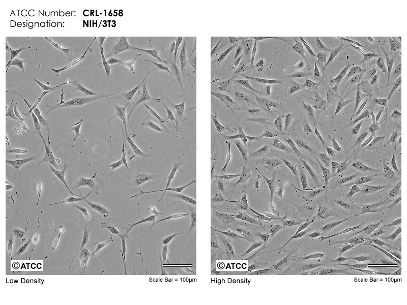
Mouse emryonic fibroblast cell line (NIH/3T3). Source: ATCC
Cell 2 is a neuronal cell. It is a hybrid cell line derived from mouse neuroblastoma cells and rat glioma cells. An example cell line is NG108-15, pictured below.
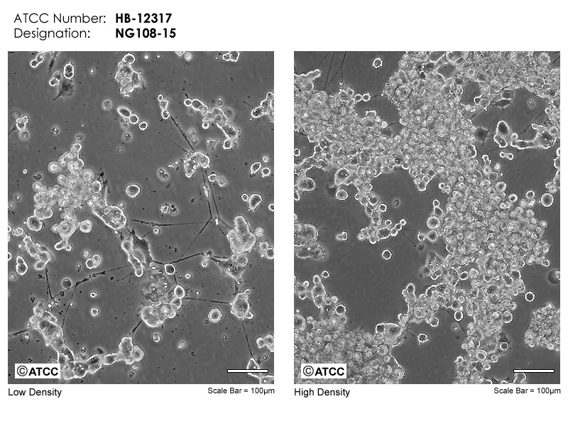
Mouse/rat neuronal cell line (NG108-15). Source: ATCC
Cell 3 an osteoblast cell from a human. An example cell line is U-2 OS, which is a cell line derived from human osteosarcoma (bone cancer) cells. These cells are pictured below.
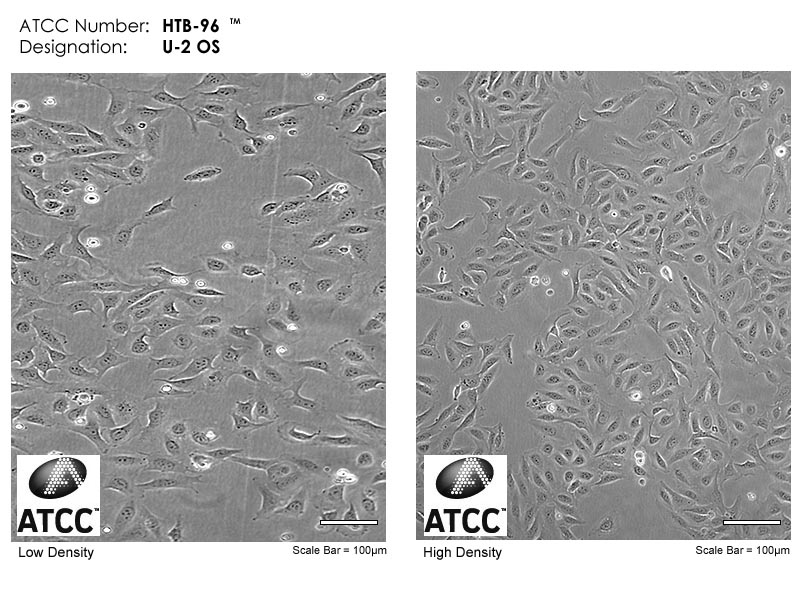
Human osteoblast cell line (U-2 OS). Source: ATCC
Cell 4 is an epithelial cell from a hamster. An example cell line is CHO-K1, pictured below.
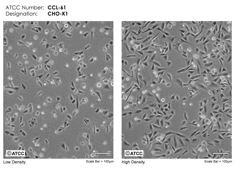
Hamster epithelial cell line (CHO-K1). Source: ATCC
Sources
(1) https://doi.org/10.1016/j.cub.2012.12.015
(2) https://www.nature.com/articles/s41580-019-0172-9(3) https://www.nature.com/articles/s41580-021-00366-6?fromPaywallRec=false
(4) https://www.mbi.nus.edu.sg/mbinfo/what-is-cell-migration/
(5) https://www.ncbi.nlm.nih.gov/pmc/articles/PMC2542983/
(6) https://www.news-medical.net/health/Formation-of-Gastrula.aspx
(7) https://www.animal-reproduction.org/article/5b5a6087f7783717068b47f5
(8) https://pubmed.ncbi.nlm.nih.gov/32119281/
(9) https://www.sciencedirect.com/science/article/pii/B9780128184226000095
(10) https://www.ncbi.nlm.nih.gov/pmc/articles/PMC10182861/
(11) https://www.ncbi.nlm.nih.gov/pmc/articles/PMC7615151/
(12) https://www.ncbi.nlm.nih.gov/books/NBK545263/
(13) https://www.ncbi.nlm.nih.gov/pmc/articles/PMC7576879/
(14) https://www.ncbi.nlm.nih.gov/pmc/articles/PMC8285443/
(16) https://www.nature.com/articles/s41416-020-01149-0
(17) https://www.ncbi.nlm.nih.gov/pmc/articles/PMC3628536/